

You notice a new red mark on your cheek while getting ready for work, or a delicate blue web behind your knee when you change clothes. It may look harmless, but the name vascular lesion can sound more alarming than the mark itself. Many people aren't sure whether they should book a GP appointment, a skin check, or an aesthetic consultation.
The answer depends on what the mark is, how long it has been there, and whether it's changing or causing symptoms. This guide explains what vascular lesions are, how common types differ, which warning signs need medical review, and where treatments such as sclerotherapy, IPL and 3D Vasculase laser may fit.
Table of Contents
- What Vascular Lesions Actually Are
- Common Types You Can See on the Skin
- What Causes Vascular Lesions and Who Gets Them
- Symptoms, Diagnosis and Red Flags to Watch For
- Treatment Options from Sclerotherapy to 3D Vasculase
- What to Expect During and After Laser Treatment
- Vascular Lesion FAQs for Clinic Patients
What Vascular Lesions Actually Are
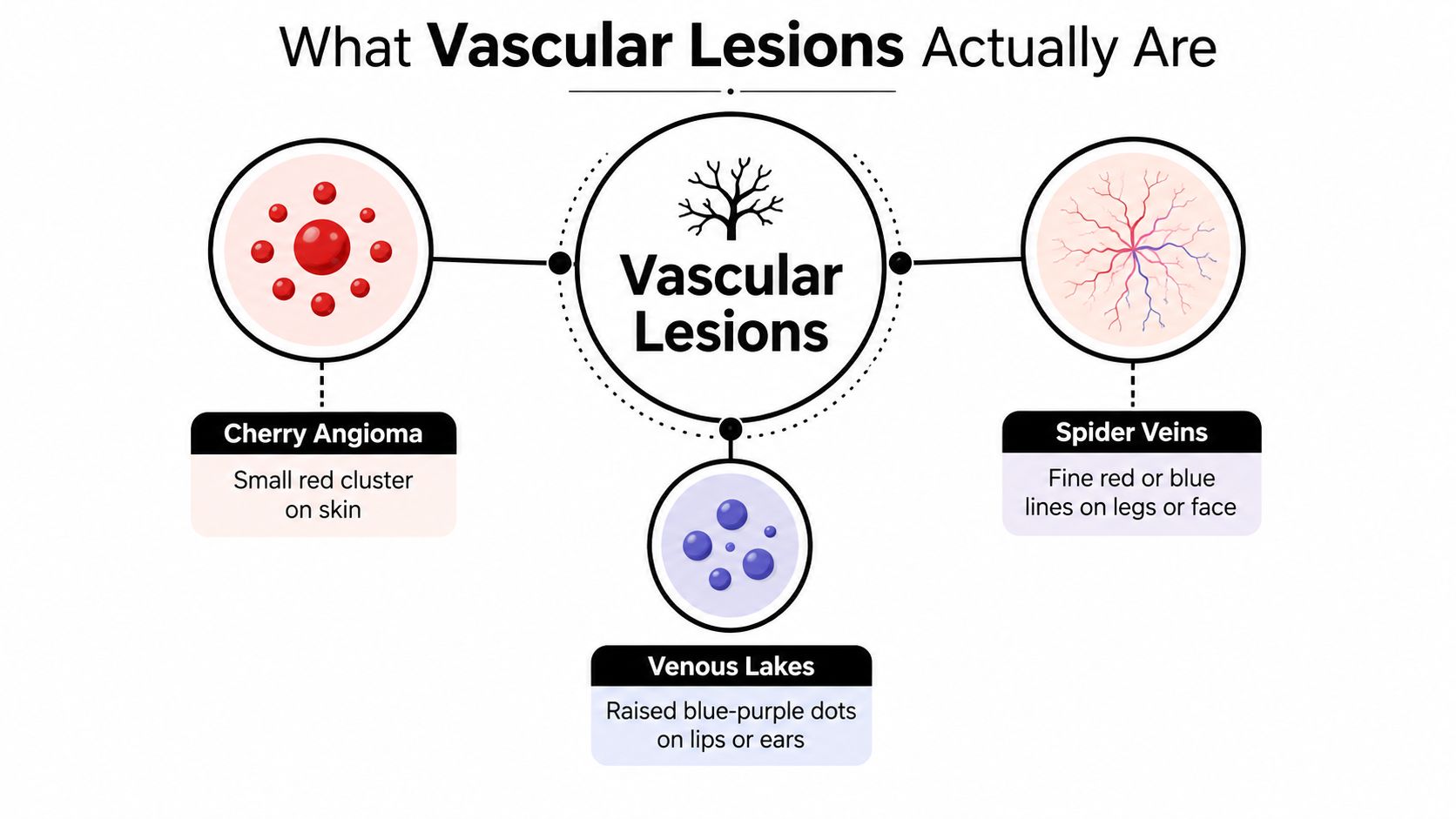
Think of the blood vessels in your skin as a fine road network on a map. Capillaries are the tiny side streets, veins are larger routes carrying blood back through the body, and lymphatic vessels form a separate drainage network. A vascular lesion appears when part of that network becomes widened, clustered, unusually formed or more visible than the surrounding skin.
That definition covers a broad range of marks. A single thread vein, a small cherry angioma, a port-wine stain and a complex venous malformation can all fall under the wider term, although they aren't the same condition and shouldn't automatically receive the same treatment. Vascular lesions also differ from pigmentation, freckles and moles, which are mainly related to melanin or changes in skin cells rather than visible blood vessels.
Most superficial lesions are harmless and are treated because they're noticeable, persistent or frustrating. Some are present from birth, while others develop gradually with age, sun exposure, hormonal changes or repeated pressure on the vessels. UK specialist sources describe vascular anomalies as a broad group of congenital and acquired lesions, ranging from simple discolouration to large, complex abnormalities. A specialist review states that vascular anomalies affect approximately 1 in 22 children, with around 60% found in the head and neck region (BAPRAS guidance on vascular anomalies).
The key distinction
A flat red mark may involve capillaries. A blue, soft or compressible mark may involve veins. A deeper swelling, warmth, pulsing or rapid change can suggest a more complicated vascular problem. Those differences matter because a laser designed for small superficial vessels isn't a substitute for assessment of a deeper malformation.
If you're organising a clinic appointment, practical preparation resources such as the Repose Healthcare basket overview may help you review what you need before attending. The important first step, however, is identifying the lesion correctly rather than choosing a treatment based only on colour.
Common Types You Can See on the Skin
Vascular lesions can look quite different in the mirror. The most useful clues are colour, texture, shape, location and timing. A mark that's been present since birth raises different questions from a red dot that appeared gradually in adulthood.
Thread veins and spider naevi
Thread veins are fine red, purple or blue lines visible beneath the skin. They can appear as isolated lines or spread from a central point like tiny tree branches. On the face, they often sit around the nose and cheeks. On the legs, they may form small networks behind the knees or around the thighs.
A spider naevus has a similar branching pattern, often with a central red point and delicate vessels radiating outwards. These lesions are usually acquired rather than present from birth, although individual causes vary.
Cherry angiomas
A cherry angioma is usually a small, bright red mark that may be flat or slightly domed. Many people compare it to a pinprick drop of jam. These lesions commonly appear on the trunk, arms or other areas of the body and tend to become more noticeable with age.
Because a cherry angioma is often raised, it can catch on clothing or bleed if scratched. A changing or repeatedly bleeding red spot still deserves assessment rather than being assumed to be a cherry angioma.
Port-wine stains
A port-wine stain is a flat pink, red or wine-coloured patch that is generally present from birth. It can look like colour has soaked into the skin, rather than sitting on the surface. Port-wine stains may occur on the face or body and can become darker or thicker over time.
They belong to the capillary malformation group. Their long-term assessment may differ from that of acquired facial thread veins, particularly when the mark is extensive or associated with other symptoms.
Infantile haemangiomas
An infantile haemangioma, sometimes called a strawberry mark, is a raised, bright red growth that typically becomes visible during the early weeks of life. It may grow for a period before gradually changing. Babies with larger, rapidly growing or strategically located lesions need professional assessment because location and size can affect function.
Venous lakes and broken capillaries
Venous lakes are soft blue or purple marks that commonly occur on the lips or ears. Broken capillaries, meanwhile, look like fine red lines or small patches across the cheeks and nose. Facial redness may also occur with rosacea, so a consultation should consider the broader skin pattern rather than treating every red area as an isolated vessel.
| Lesion Type | Typical Appearance | Common Location | Usually |
|---|---|---|---|
| Thread veins | Fine red, purple or blue lines | Face and legs | Acquired |
| Spider naevi | Branching vessels from a central point | Face, neck and upper body | Acquired |
| Cherry angiomas | Bright red flat or domed dots | Trunk and arms | Develop with age |
| Port-wine stains | Flat pink, red or wine-coloured patch | Face or body | Present from birth |
| Infantile haemangiomas | Raised bright red “strawberry” growth | Various sites in infants | Develop early in life |
| Venous lakes | Soft blue-purple spot or bump | Lips and ears | Acquired |
| Broken capillaries | Fine red vessels or diffuse redness | Cheeks and nose | Acquired |
For a clinic-specific explanation of facial vessels, see the broken capillaries treatment information.
What Causes Vascular Lesions and Who Gets Them
A vascular lesion can begin before birth or become visible later. The cause depends on how the blood or lymph vessels developed, and on changes affecting the skin and vessel walls over time.
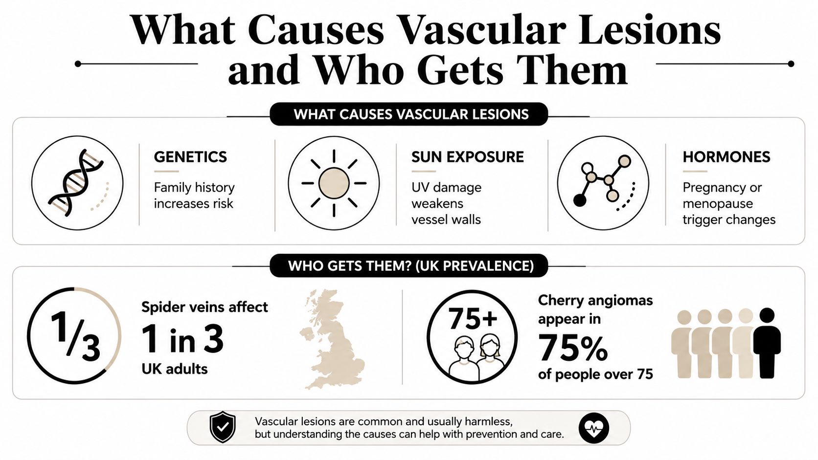
Congenital and developmental factors
Port-wine stains and many vascular malformations are present from birth. Infantile haemangiomas usually become noticeable during early infancy, even when they are not obvious at delivery. Developmental differences may involve capillary, venous, lymphatic or arterial vessels, so a lesion can sit near the skin surface or deeper within the tissue.
The vessel type matters because each behaves differently. Venous malformations are described as the most common type of vascular anomaly by UK specialist information. A British interventional radiology review describes vascular malformations as rare, occurring at approximately 1 in 7,000 births (Leeds Vascular Anomalies Service definitions).
Acquired influences
Sun exposure can make small facial vessels easier to see, especially across the cheeks and around the nose. Rosacea may cause persistent flushing and dilated capillaries. Hormonal shifts during pregnancy or menopause can also affect vascular visibility, while prolonged standing may make leg veins more prominent.
Skin changes with age. As it becomes thinner and superficial vessel walls lose resilience, marks that once went unnoticed may become clearer.
What changes your likelihood
Fairer skin can make red or blue vessels more visible, although vascular lesions occur across all skin tones. Family history may influence susceptibility. Work or daily routines involving heat or long periods of standing can also increase the tendency for visible leg vessels.
These conditions are also seen in paediatric care. A UK and European review reports that vascular anomalies affect approximately 1 in 22 children, and a separate review notes that around 60% occur in the head and neck region (head and neck vascular anomaly review). Those figures do not mean every red mark indicates a serious condition. They explain why specialist assessment begins with classification, rather than treating every vascular lesion as a cosmetic concern.
Symptoms, Diagnosis and Red Flags to Watch For
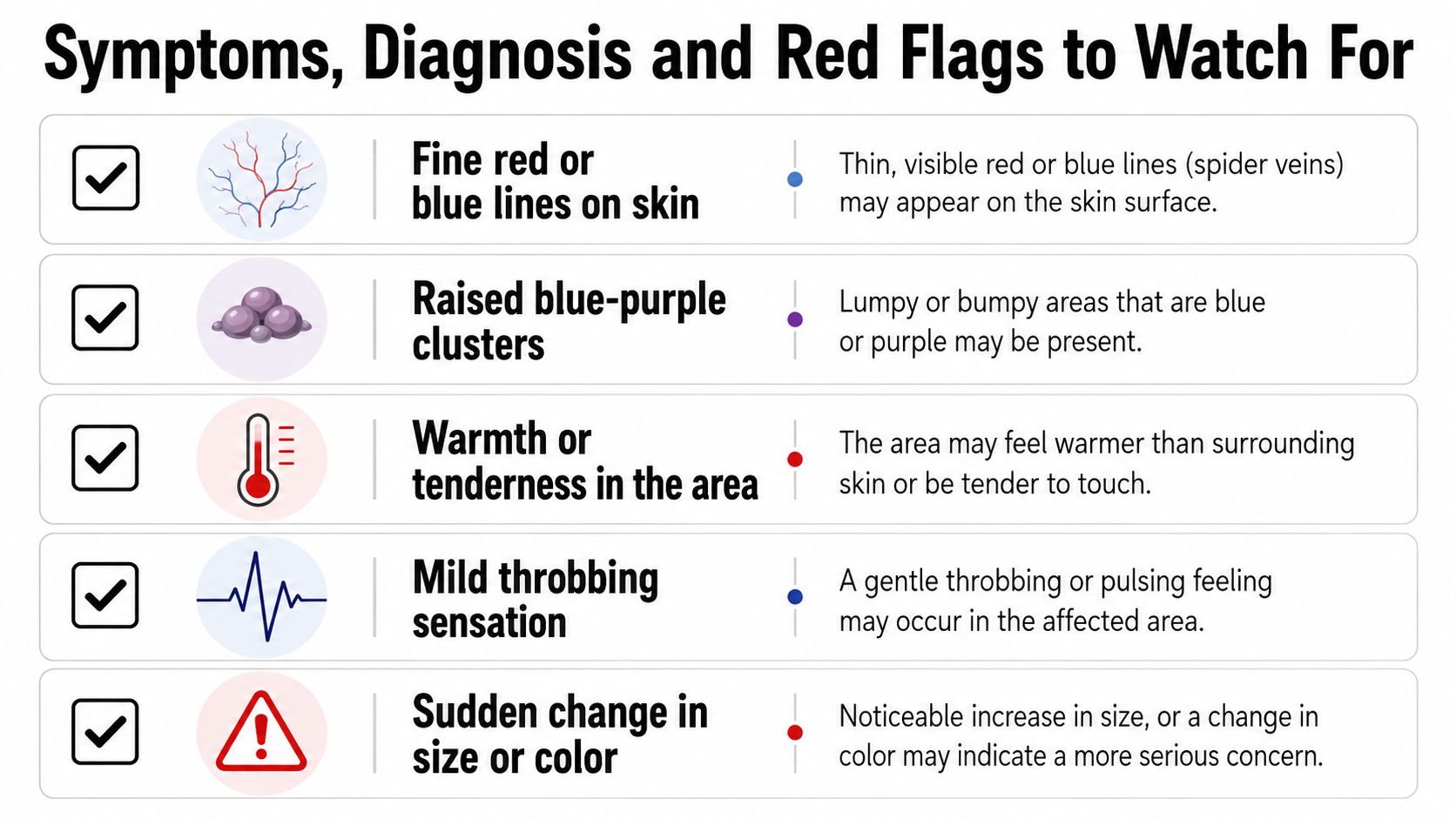
A vascular lesion may be purely visual, or it may produce physical sensations. Patients commonly describe fine red or blue lines, a raised purple cluster, local warmth, tenderness or a mild throbbing feeling. The symptom pattern helps a practitioner decide whether the concern is likely to be superficial and cosmetic or whether it needs medical investigation.
What happens in a consultation
A responsible consultation begins with questions. The practitioner may ask when the mark appeared, whether it has changed, whether it bleeds, and whether you have pain, swelling or a relevant medical history. They'll inspect the colour, borders, surface and depth, sometimes using magnification or dermoscopy to assess the skin more closely.
Ultrasound may be appropriate when a lesion appears deeper, compressible or connected to larger veins. Low-flow venous or lymphatic malformations can require a different pathway from small facial telangiectasia. NHS information explains that some low-flow malformations need no treatment unless they cause pain or functional limitation, while specialist services may use imaging and multidisciplinary assessment for more complex cases.
Red flags that change the plan
Book a GP review before an aesthetic procedure if a lesion:
- Grows quickly: A sudden increase in size needs medical assessment.
- Bleeds repeatedly: Don't assume recurring bleeding is just from friction.
- Swells suddenly: Rapid swelling, marked pain or heat may require prompt attention.
- Changes asymmetrically: An uneven change in outline or structure should be examined.
- Alters in colour or surface: New pigmentation, crusting or ulceration shouldn't be treated as a routine cosmetic vessel.
Safety rule: A laser consultation should confirm that the mark is suitable for aesthetic treatment. It shouldn't replace medical assessment of a new or changing lesion.
If the lesion is associated with limb swelling, persistent pain, functional difficulty or signs of a deeper abnormality, ask your GP about an appropriate specialist referral. A superficial thread vein and an arteriovenous malformation may both look “vascular”, but they aren't interchangeable.
Treatment Options from Sclerotherapy to 3D Vasculase
Treatment starts with the vessel type and location, not with the machine. A small facial telangiectasia may respond well to a precise laser, while larger leg veins may be better suited to an injection-based approach. Some asymptomatic malformations need observation rather than intervention.
Sclerotherapy
Sclerotherapy involves injecting a sclerosing solution into selected veins. The solution irritates the vessel lining, encouraging the vein to close so the body can gradually clear it. It's commonly considered for larger reticular or varicose leg veins, and the injection plan depends on vessel size, depth and circulation.
NHS treatment information describes embolosclerotherapy as a way to inflame and close a malformation, reducing symptoms such as pain, swelling and bleeding. Follow-up is commonly arranged 8 to 10 weeks after treatment according to an NHS leaflet cited in the UK specialist review (UK vascular anomaly treatment review).
IPL
Intense pulsed light, or IPL, uses a broad spectrum of light rather than one single laser wavelength. It may be selected for diffuse facial redness and small telangiectasia spread across an area. Because the energy is broader, the practitioner must match settings carefully to the skin tone, redness pattern and treatment goal.
Dedicated vascular laser treatment
A dedicated vascular laser uses a selected wavelength to heat blood vessels more precisely. 3D Vasculase uses a 980nm diode laser, targeting haemoglobin in superficial vessels. Its 0.2 to 0.5mm spot size is designed for detailed work on thread veins and facial telangiectasia, where accuracy matters more than treating a large area with bulk heat.
Practitioners may discuss one to three sessions, usually separated by four to six weeks, depending on the number, depth and response of the vessels. The treatment is described for Fitzpatrick skin types I to IV, with suitability confirmed during consultation rather than assumed from a website description. You can read more about the 3D Vasculase vascular laser treatment before booking.
| Treatment | Best For | Mechanism | Typical Sessions | Downtime |
|---|---|---|---|---|
| Sclerotherapy | Larger leg veins and selected malformations | Injected solution closes the vessel | Individual plan | Depends on area and response |
| IPL | Diffuse redness and small surface vessels | Broad-spectrum light heats vascular targets | Individual plan | Usually limited, but varies |
| 3D Vasculase | Thread veins and facial telangiectasia | 980nm diode laser targets haemoglobin | One to three may be discussed | Usually low, subject to treatment response |
| Other vascular lasers | Selected superficial lesions | Wavelength-specific vessel heating | Individual plan | Varies by device and lesion |
A trained practitioner should explain why one route fits your lesion and why another doesn't. “More powerful” isn't automatically better when the target is a delicate superficial vessel.
What to Expect During and After Laser Treatment
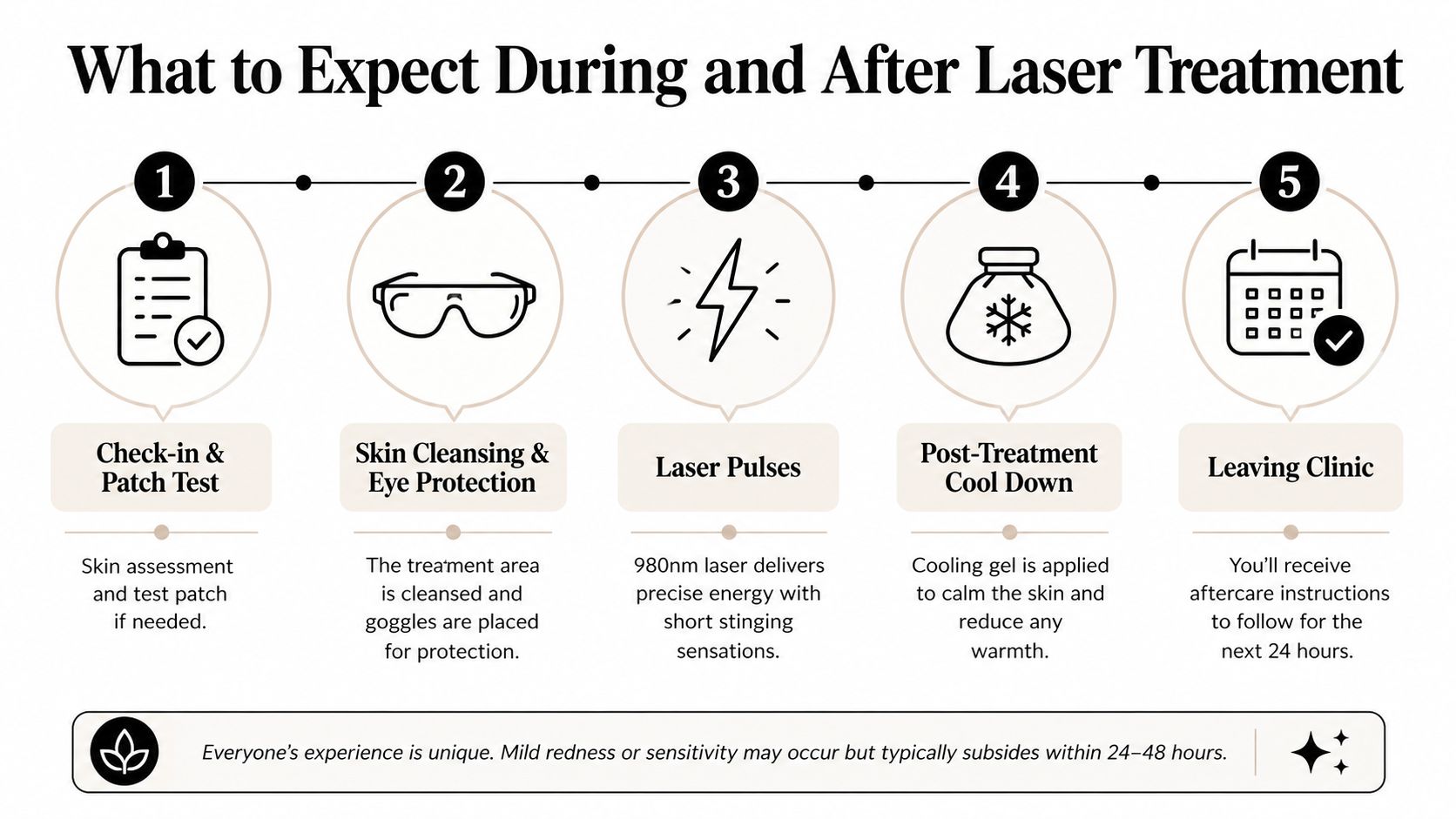
A typical appointment begins with a review of your medical history, skin condition and treatment area. The practitioner may recommend a test patch when needed, particularly if they need to assess how your skin responds before treating a larger area.
The skin is cleansed, photographs may be taken for comparison, and protective eyewear is fitted. With 3D Vasculase, the 980nm laser delivers short pulses that can feel like a quick sting or snap. The treated vessel may darken as the blood inside it coagulates, which can look more noticeable immediately after the session.
The fading process
Don't judge the final result in the treatment room. The body needs time to clear the treated vessels, and visible veins may fade over four to twelve weeks. A follow-up review at six weeks can help the practitioner assess progress and decide whether another session is appropriate.
You may notice temporary redness, mild swelling, warmth, tenderness or small areas of darkening. These effects should settle as the skin recovers. Contact the clinic if you develop worsening pain, extensive blistering, spreading swelling, signs of infection or a reaction that concerns you.
For area-specific guidance, review the information on treatments for spider veins on the face.
Protecting the treated skin
Follow the clinic's instructions closely:
- Use SPF 50: Protect the area from ultraviolet exposure while it heals.
- Keep cleansing gentle: Avoid scrubbing, picking or abrasive products.
- Pause retinoids on the site: Restart only when your practitioner says the skin barrier has recovered.
- Avoid heat and exertion: Skip saunas, alcohol and strenuous exercise for 48 hours.
- Cool the area carefully: Use the recommended cooling product or compress without applying harsh pressure.
The aim is simple. Reduce irritation, protect the skin from extra pigment changes and give the treated vessels time to resolve.
Vascular Lesion FAQs for Clinic Patients
Does laser treatment hurt?
Most patients describe a short stinging, snapping or warming sensation rather than sustained pain. If you use a 0 to 10 pain scale, think of the experience as a brief, localised sensation that varies with the vessel, body area and personal tolerance. Tell the practitioner if the heat feels excessive, because comfort and skin safety should be monitored throughout the appointment.
How much does treatment cost?
There isn't one responsible price for every vascular lesion. The quote depends on the size of the area, number of vessels, body location and likely number of sessions. A consultation gives you a more useful estimate than a generic menu price because the practitioner can decide whether laser, IPL, sclerotherapy or medical referral is appropriate.
Can I combine vascular treatment with other procedures?
Sometimes, but sequencing matters. Skin rejuvenation, microneedling, anti-wrinkle injections and laser vascular work may all be available within the same clinic, yet they shouldn't automatically be performed together. Your practitioner may separate treatments to reduce irritation and allow the skin to recover, with the waiting period based on the procedure, treatment area and your healing response.
Is 3D Vasculase suitable for every skin tone?
The treatment is described for Fitzpatrick skin types I to IV, but a skin-type label alone doesn't guarantee suitability. Recent tanning, active inflammation, medication, pigmentation risk and the lesion's depth also matter. A consultation and test patch, where appropriate, help the practitioner choose safe settings or recommend a different route.
Are the results permanent?
A treated vessel may remain reduced or cleared, but your skin can develop new vessels later. Sun exposure, ageing, hormonal changes and the original tendency toward visible veins can all influence maintenance needs. Think of treatment as addressing the vessels that are present now, not as a guarantee that no new lesion will ever appear.
What should I look for in a UK clinic?
Choose a clinic that completes a proper consultation, checks medical history, explains contraindications and knows when to refer you to a GP or specialist service. Ask who performs the treatment, what training they hold, how the device is used, what aftercare is provided and how the clinic handles complications. Clear answers are more important than pressure to book quickly.
3D Aesthetics Leamington Spa offers consultation-led vascular lesion treatment using 3D Vasculase, alongside other non-surgical skin and facial services. Visit 3D Aesthetics Leamington Spa to discuss your mark with a trained therapist, confirm whether laser treatment is suitable, and receive a personalised plan for your skin and goals.
← Back to Home