
You've looked in the mirror before a consultation and noticed something you couldn't quite describe. One cheek seems slightly flatter, your jawline appears different from one side to the other, or a treatment area looks improved in person but unchanged in a photograph. That uncertainty is common because a mirror and a flat photograph offer only a partial view of a three-dimensional face.
3D facial scanning gives your practitioner another way to assess those changes. It creates a digital surface map that records facial contours, depth, volume and asymmetry, helping turn a subjective visual impression into a measurable baseline. For anyone considering non-surgical facial rejuvenation, that can make treatment planning and follow-up easier to understand.
Table of Contents
- Understanding 3D Facial Scanning Technology
- Clinical Benefits and Precision Treatment Planning
- Comparing 3D Scans with Traditional 2D Photography
- Your Scanning Experience at 3D Aesthetics Leamington Spa
- Common Aesthetic Use Cases and Applications
- Navigating Data Privacy and Biometric Concerns
- Frequently Asked Questions About Facial Scanning
Understanding 3D Facial Scanning Technology
A client may see a flatter cheek in the mirror, while a photograph makes the difference seem larger or smaller. Expression, lighting, camera position and lens choice all affect that impression. A mirror offers a changing reflection, and a 2D image compresses a three-dimensional face into a flat record.
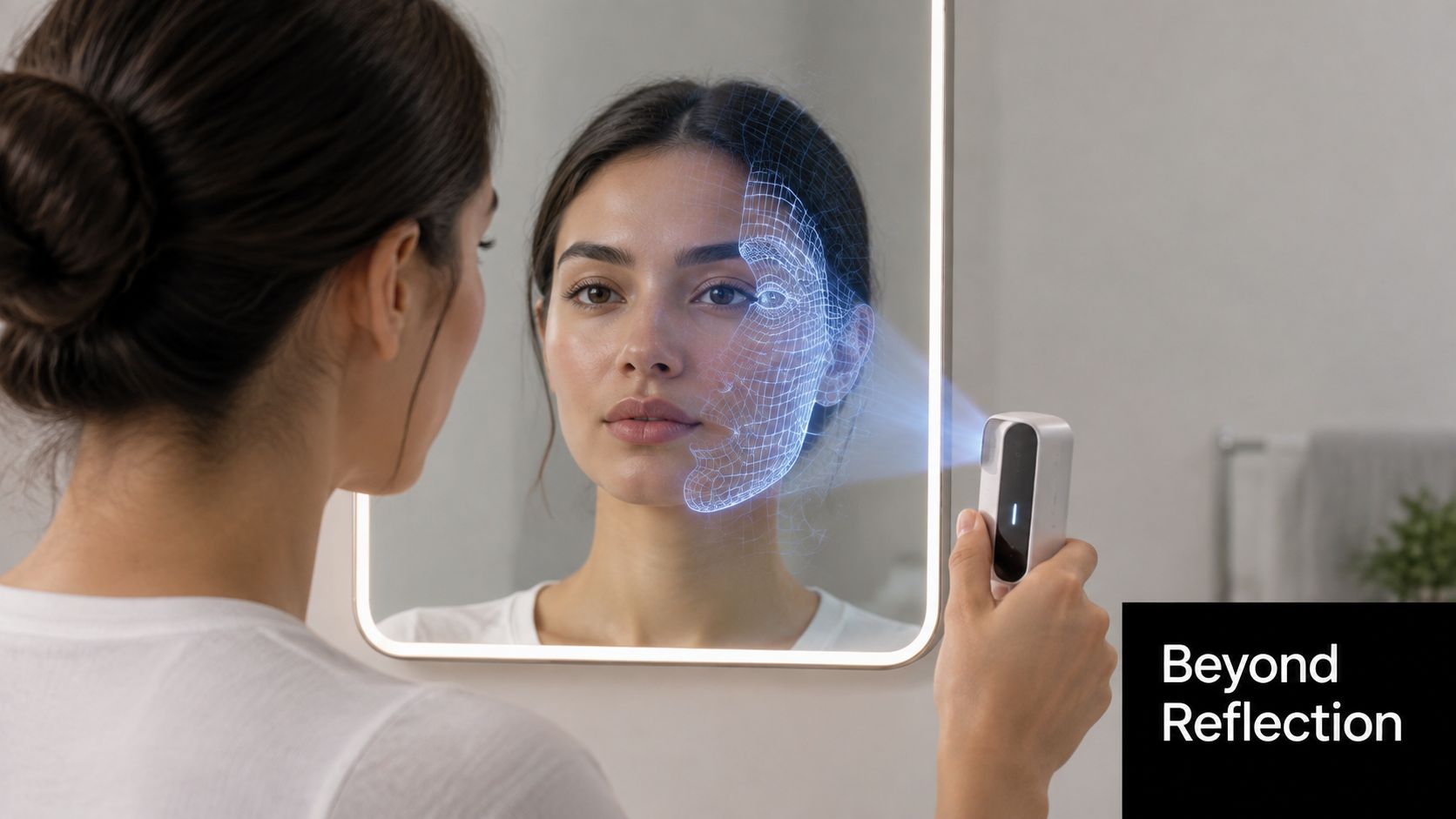
3D facial scanning works differently. A handheld or fixed scanner uses imaging sensors, often structured light or infrared, to capture the face's surface geometry. Software combines those readings into a digital mesh, creating a map of depth and shape rather than relying only on colour and shadow.
What the scan records
The model can show:
- Surface contours, including the curves of the cheeks, lips, chin and forehead.
- Relative depth, which helps identify depressions, projection and changes in facial profile.
- Asymmetry, allowing corresponding areas on the left and right sides to be compared.
- Volume changes, which can be reviewed against a later scan captured under comparable conditions.
The result is closer to a digital measuring surface than a facial-recognition image. In an aesthetic consultation, its purpose is generally to map the treatment area and support clinical discussion. The information collected, stored and retained depends on the scanner, software and clinic's data practices, so ask who controls the files, how long they are kept and whether they are deleted on request before consenting.
Academic work at the University of York built one of the UK's largest structured 3D facial image datasets. The collection described in the UK government's biometrics report contains around 5,000 3D facial images from approximately 350 people, with about 15 scans per model, supporting research into recognition, reconstruction and measurement (UK Biometrics and Surveillance Camera Commissioner Annual Report). A UK forensic facial reconstruction paper from 1997 also recorded 3D reconstruction methods in scientific practice.
Practical rule: Use the scan as a measurement aid, alongside consultation, examination and medical history.
The underlying anatomy helps explain the images. Muscles, soft-tissue volume and bone structure can all influence the surface you see, as shown in this 3D muscle model guide. Similar mapping principles can assess other areas of the body, including the measurement methods described in this guide to body measurement tracking.
Clinical Benefits and Precision Treatment Planning
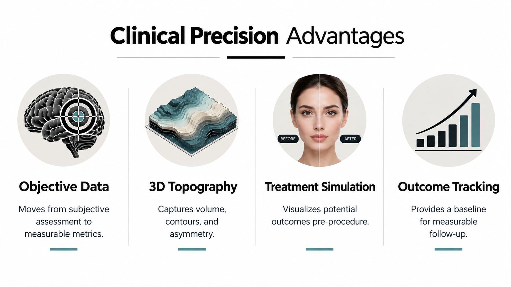
The value of a facial scan isn't just that it produces an impressive rotating image. Its clinical usefulness comes from giving the practitioner a repeatable reference point before treatment, then allowing later images to be compared with that baseline.
A consultation still begins with conversation and observation. You explain what concerns you, the practitioner examines your skin and facial movement, and together you discuss realistic outcomes. The 3D model adds another layer by showing how those concerns appear across the face as a whole.
Why precision changes the conversation
Facial aesthetics often involves subtle change. A small improvement in cheek projection, a gradual reduction in lower-face laxity or a difference in scar contour can be difficult to judge from memory. A digital model lets the practitioner review the same anatomical region from different angles and discuss the proposed treatment in a more concrete way.
UK craniofacial research from Cardiff found that repeated low-cost 3D facial scans had differences of less than 1%, while the study also cited prior clinical scanner accuracy in the 0.2 to 1.0 mm range (Cardiff craniofacial research). That doesn't mean every scan is automatically perfect. Positioning, facial movement, hair, lighting and the device itself all matter. It does show why a properly calibrated system can be suitable for monitoring small contour or volume changes over time.
A separate UCL comparison illustrates why equipment selection matters. Against a reference 3dMDface system, one portable structured-light scanner recorded 90% of data points within 2 mm and an RMS error of 0.71 mm ± 0.28 mm. Another lower-cost system recorded 80% within 2 mm and an RMS error of 1.33 mm ± 0.46 mm (UCL facial imaging comparison).
What this means for treatment planning
A practitioner can use the scan to:
- Establish a baseline, recording the starting contours before an intervention.
- Assess asymmetry, so the plan reflects your anatomy rather than an assumed ideal.
- Discuss proportion, including how the cheeks, lips, chin and jaw relate visually.
- Review progress, comparing later scans when treatment response needs objective assessment.
For dermal fillers, the model may support discussion of where restoring volume could improve balance, although it can't determine the correct product, dose or injection technique on its own. For skin-tightening treatments, it can help document the starting degree of laxity and provide a visual reference for follow-up. For scar work, surface mapping can help distinguish a shallow textural change from a more pronounced contour irregularity.
The most useful result is often better communication. You and your practitioner can look at the same image, identify the same region and agree on what improvement means before treatment begins.
Comparing 3D Scans with Traditional 2D Photography
Traditional clinical photography remains useful. It can document colour, redness, pigmentation, bruising and visible skin texture, particularly when the camera, lighting and patient position are kept consistent. The limitation is that a flat photograph compresses a three-dimensional face into a single plane.
Manual measurements have a similar problem. A practitioner can measure distances between landmarks, but a few linear measurements can't describe the full curve of a cheek, the depth of a hollow or the transition between the chin and neck. A 3D model adds surface shape to the assessment.
| Feature | Traditional 2D Photography | 3D Facial Scanning |
|---|---|---|
| Depth | Implied through shadows and perspective | Recorded as part of the facial surface map |
| Volume | Difficult to judge reliably from a flat image | Can be assessed through mapped contours and comparative models |
| Asymmetry | Visible from selected angles | Reviewed across the digital face from multiple viewpoints |
| Lighting dependence | Strong influence from shadows, exposure and colour balance | Shape data can provide a clearer topographical reference, although scan conditions still matter |
| Follow-up comparisons | Useful when views and camera settings are carefully standardised | Allows digital comparison of corresponding facial surfaces |
| Skin colour and texture | Often clearer and more familiar | May depend on the scanner's colour-capture capability |
| Patient communication | Easy to understand but limited to fixed views | Rotatable model can make contours and treatment areas easier to discuss |
| Best role | Documenting appearance and skin changes | Mapping shape, depth, contour and measurable change |
Where the difference matters
Cheek hollowing is a good example. In a front-facing photograph, a shadow may make a hollow appear deeper than it is, while bright lighting may soften it. A side view can show projection, but it still won't capture the complete transition from the cheek to the temple and lower face.
Jawline definition presents another challenge. A photograph can show the outline, but it can't fully describe whether an apparent lack of definition relates to skin laxity, soft-tissue volume, chin projection or the angle of capture. A 3D view gives the practitioner more context before suggesting a treatment pathway.
A photograph records how your face looked at one moment. A well-controlled 3D scan helps describe how its surfaces are shaped.
Neither method should be treated as a standalone diagnosis. The strongest consultation combines clinical examination, photographs, patient goals and appropriate imaging. If a clinic uses 3D scanning, ask whether follow-up scans will reproduce the same position and conditions. Consistency matters as much as sophistication.
Your Scanning Experience at 3D Aesthetics Leamington Spa
A facial scan during a consultation is designed to be straightforward. You'll usually sit or stand in a neutral position while the practitioner asks you to relax your face and keep still. The scanner is moved around the treatment area, or you'll face a fixed imaging system, depending on the equipment used.
There's no incision and no contact with the skin during the imaging itself. The process is non-invasive, painless and radiation-free. The practitioner may ask you to remove glasses, move hair away from the face and keep your lips in a neutral position so that the software can capture the relevant landmarks more consistently.
What you'll see on screen
The scan produces a three-dimensional representation that can be rotated. Your practitioner may use colour shading, heat maps or contour lines to show differences in surface position. These colours aren't a universal medical grading system, so their meaning depends on the software and the comparison being displayed.
A heat map may show where one scan sits higher, lower or differently from another. Contour lines can help illustrate the shape of a cheek, the edge of a scar or the profile beneath the chin. Ask the therapist to explain the scale before drawing conclusions from the colours.
The consultation should connect the image to your goals. For example, if you're concerned about lower-face laxity, the practitioner might review the jawline and neck from several angles. If you're considering resurfacing, the discussion may focus more on skin texture, pigmentation and scar visibility, areas where photographs can still provide important information.
Questions worth asking
Before agreeing to treatment, ask:
- What is being measured? Clarify whether the scan maps surface shape, colour, volume comparisons or another feature.
- Will the scan be repeated? Find out whether future images can be compared with the original baseline.
- Who can access the file? Ask which staff, software providers or external parties may handle it.
- How long is it retained? Request a plain-English explanation of storage and deletion arrangements.
- What happens if the scan and examination differ? Your practitioner should explain how clinical judgement takes priority.
A clinic's approach to scanning should feel as organised as its wider consultation process. If you're also researching other non-invasive procedures, understanding each stage of a laser hair removal process can help you see how preparation, treatment delivery and follow-up fit together.
Common Aesthetic Use Cases and Applications
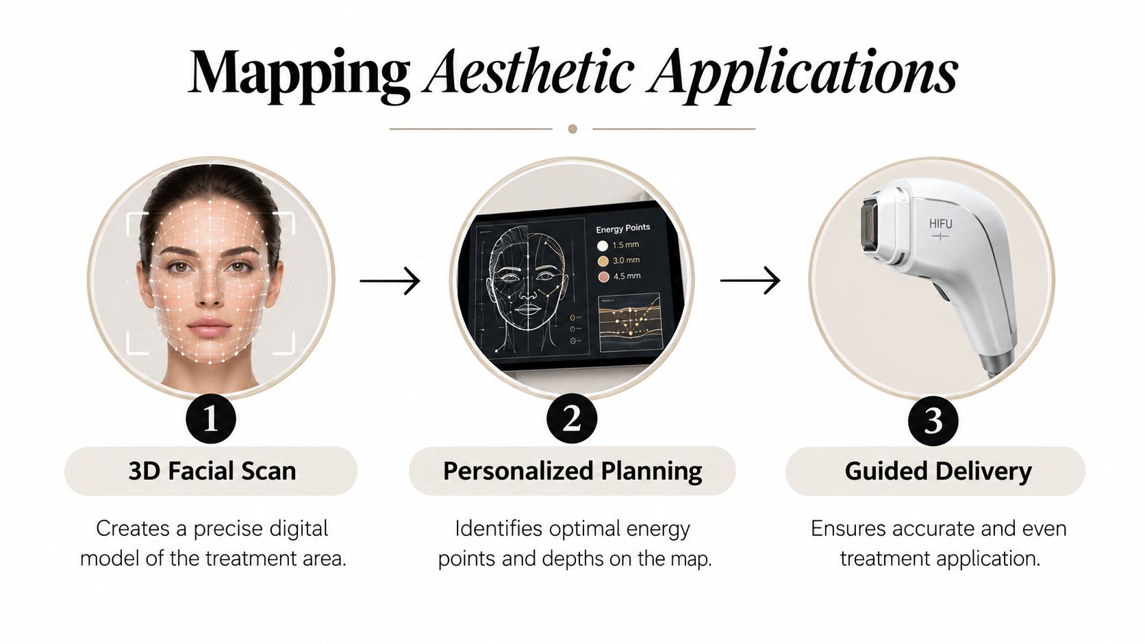
A 3D scan is most useful when it changes what the practitioner does next. It isn't a treatment by itself. It acts as a planning reference that can help match a procedure to the location, depth and character of the concern being assessed.
HIFU and radio-frequency tightening
For HIFU facelift planning, facial mapping can help organise the conversation around areas of laxity and the relationship between the lower face, jawline and neck. It doesn't replace the practitioner's knowledge of facial anatomy or the device protocol. Instead, it creates a visual record of the starting contour and helps identify where treatment should be considered carefully.
Radio-frequency treatments can be discussed in a similar way. A scan may document the shape of the treatment area before a course and support a more consistent review afterwards. The practitioner still needs to assess skin quality, contraindications, comfort and whether the treatment is suitable.
Microneedling, resurfacing and scars
Acne scarring isn't always a flat, uniform problem. Some scars are shallow depressions, some have sharper edges, and some sit alongside pigmentation or enlarged pores. A surface map can help show the contour component, while clinical photographs and examination remain important for colour and texture.
For 3D Dermaforce microneedling or fractional CO2 resurfacing, this information can support a more targeted discussion about which areas need attention and what a realistic improvement may look like. It can also help separate a change in surface depth from a change in redness, since those features require different forms of assessment.
Fillers and facial balance
Dermal filler planning benefits from seeing the face as a connected structure. A practitioner may assess the relationship between the cheek, under-eye area, lips, chin and jawline rather than treating one feature in isolation. A rotatable model can make that conversation easier, particularly when a small imbalance is more noticeable from an oblique view than from the front.
The scan doesn't decide whether filler is appropriate. A qualified practitioner must consider your medical history, facial movement, skin condition, previous procedures and desired outcome. It can, however, provide a clearer visual framework for discussing conservative changes and avoiding a one-size-fits-all plan.
The same principle applies when tracking results. A baseline scan gives both sides a reference, but comparison is only meaningful when the face is positioned consistently and enough time has passed for the treatment's expected response to develop.
Navigating Data Privacy and Biometric Concerns
A client may look at a mirror, then review a 3D scan, and wonder whether the clinic has created a biometric record. The answer depends on what the system captures, how the software processes it and whether the information is used to identify you. Surface mapping for aesthetic planning isn't automatically the same as facial recognition for identification, but a clinic should explain that distinction clearly.
Facial-recognition processing typically involves biometric data. If it is used to identify someone uniquely, UK GDPR requires a lawful basis and an Article 9 condition. That regulatory context matters because a treatment-planning scan and a police identification system may use related imaging methods while serving very different purposes.
A 2026 Big Brother Watch briefing said the Metropolitan Police had scanned more than 1.7 million faces in London during the first part of that year, an increase of 87% compared with the same period in 2025. The briefing also said almost 800,000 people had been scanned since 2020. Cosmetic scanning has a different purpose, but this UK context explains why clients reasonably want direct answers about collection, storage and identification.
What a transparent clinic should explain
Before scanning, ask:
- Purpose: Is the file used for treatment planning and outcome review, or for another purpose?
- Identification: Does the system create a biometric template or attempt to match your identity?
- Storage: Where is the scan stored, and which provider hosts the software?
- Access: Can staff, contractors or suppliers view the data?
- Retention: When will the file be deleted, and can you request deletion where applicable?
- Consent: Can you receive treatment without agreeing to uses beyond your consultation?
A clinic should keep its privacy notice and processes current as UK data-protection requirements develop. Review its explanation of GDPR and client data before booking, and compare it with practical guidance on trust and privacy for taap.bio users. These resources cannot confirm how a particular clinic operates, so ask the provider to explain its own software, access controls and deletion process in plain language.
Frequently Asked Questions About Facial Scanning
Is the scan painful or invasive?
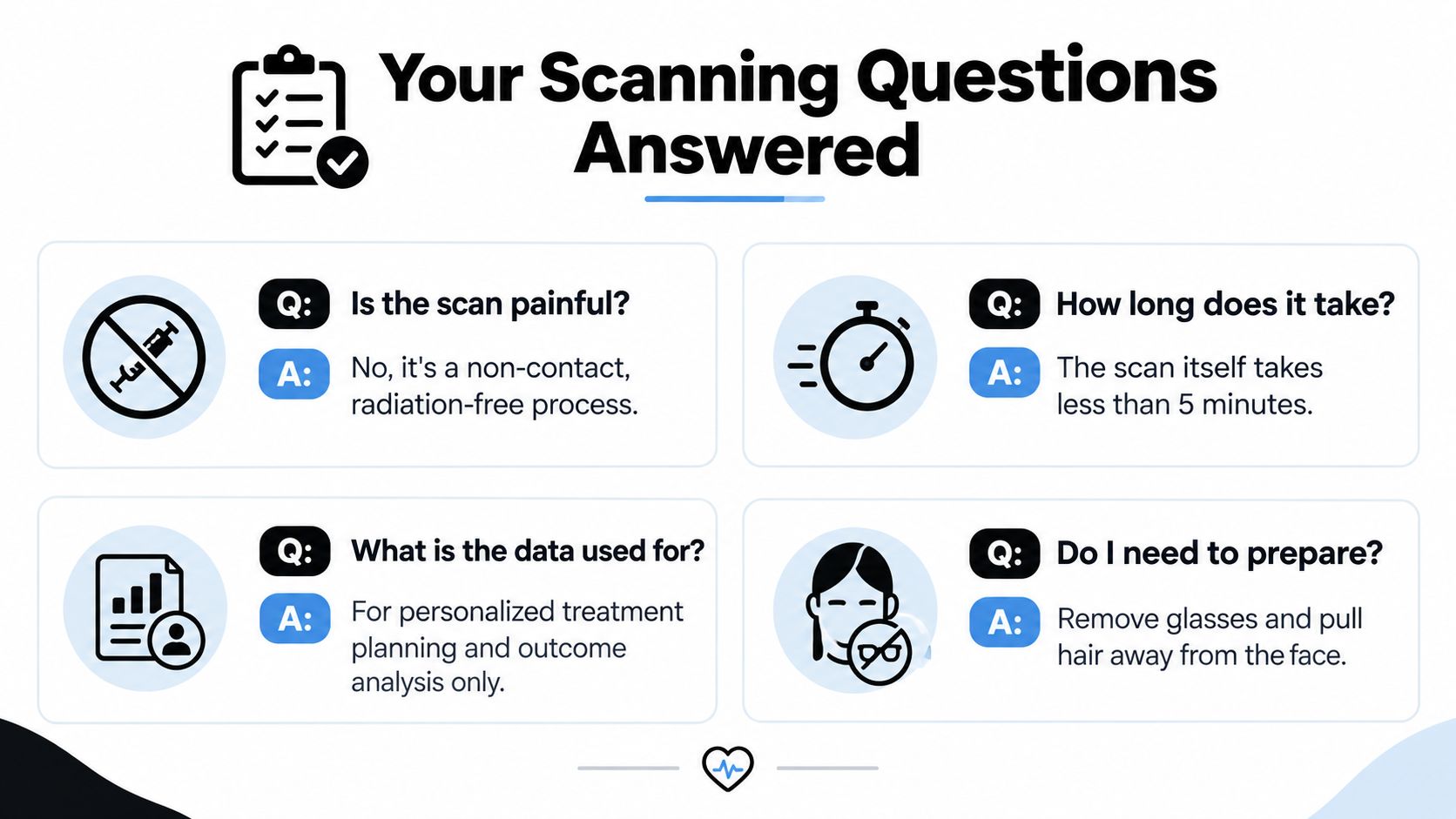
No. The imaging process is non-contact, painless and radiation-free. You'll need to remain still and follow simple positioning instructions, but the scanner shouldn't cause physical discomfort.
How long does it take?
The scan itself takes less than 5 minutes, as stated in the supplied clinic visual. Allow additional time for positioning, discussion and interpretation during the consultation. A careful consultation shouldn't be rushed just because image capture is brief.
Should I remove makeup?
Ask the clinic before attending. Heavy makeup, reflective products or anything that changes the surface may affect how the scanner reads the face, while the clinic may also want photographs under particular conditions. Arrive with glasses removed and hair pulled away from the face, as shown in the supplied preparation guidance.
Can I be scanned with a skin condition?
Often, a scan can document the surface without touching it, but suitability depends on the condition and the planned treatment. Tell the practitioner about active irritation, infection, open areas, recent procedures and prescribed products before scanning. The imaging may be safe while the proposed treatment isn't appropriate at that time.
Do metal implants cause a problem?
A facial scan doesn't use magnetic imaging, so a metal implant isn't automatically a reason you can't be scanned. You should still disclose dental work, surgical implants, injectables and previous procedures because they may affect interpretation or treatment suitability.
How often should follow-up scans happen?
There isn't one correct schedule for every treatment. The practitioner should choose timing based on the treatment's expected response, your goals and whether the scan is being used to assess contour, volume or skin change. Repeating scans too soon can create confusion if the tissue is still settling.
What does the scan prove?
It provides a structured visual and measurement reference. It doesn't prove that a treatment caused every change, predict an exact result or replace clinical judgement. The most reliable interpretation combines the scan with photographs, examination, your symptoms and the agreed treatment plan.
Can I decline storage?
Ask the clinic what options its consent process provides. You should understand whether refusing storage affects treatment, whether a temporary review is possible and how to request access or deletion. A reputable provider should answer those questions in plain English before you consent.
3D Aesthetics Leamington Spa includes facial scanning within its consultation approach to support assessment, personalised planning and outcome review for non-surgical facial treatments. Visit 3D Aesthetics Leamington Spa to arrange a consultation, ask how your scan data is handled and discuss whether 3D facial mapping suits your treatment goals.
← Back to Home