

You've checked your skin in ordinary light, then leaned towards a magnifying mirror and found that the brown patches look more noticeable than they did yesterday. Perhaps a brightening serum hasn't shifted them, or a previous peel seemed to help before the colour returned. It's frustrating, but persistent facial pigmentation isn't a sign that you haven't found a strong enough product.
The key question isn't which laser removes pigmentation fastest. It's which type of pigmentation do you have, how deep is it, and could the proposed treatment trigger more colour? A careful facial pigmentation treatment plan starts with diagnosis, then matches the least risky tool to the biology of your skin.
Table of Contents
- Why Facial Pigmentation Is Harder to Treat Than It Looks
- The Main Types of Facial Pigmentation Explained
- How a Clinician Diagnoses Your Pigmentation
- Topical Treatments and What They Actually Do
- Peels, Lasers and Light-Based Options Compared
- Who Is a Poor Candidate and What Can Make It Worse
- Planning Your Treatment and Choosing the Right Clinic
- Realistic Results, Maintenance and What Comes Next
Why Facial Pigmentation Is Harder to Treat Than It Looks
A patient may see one brown patch on the cheek. Clinical assessment can reveal several processes within it: superficial pigment, deeper pigment, inflammation left by acne, and a hormonal pattern layered over sun-related spots. The eye sees one colour, while the skin may be responding to different triggers at once.
The single most important decision before treatment is identifying the pigment's type, depth and likely cause. Epidermal pigment sits nearer the surface and may respond more readily to carefully chosen topical products, peels or pigment-selective devices. Dermal pigment lies deeper, so improvement is less predictable, and inflammation can create further discolouration. UK dermatology guidance describes dermal pigmentation as harder to treat, particularly in skin of colour, where an unsuitable device may increase treatment-induced pigmentation (Society of Cosmetic and Telemedicine Dermatology guidance).
Why a single treatment can disappoint
Melasma may reflect hormonal sensitivity, visible light, ultraviolet exposure and heat. Post-inflammatory hyperpigmentation, often called PIH, follows irritation or injury such as acne, eczema, waxing or an overly aggressive procedure. A solar lentigo, commonly called a sunspot, is a more defined area of accumulated sun-related pigment.
These conditions can overlap. Symmetrical melasma across the cheeks and forehead may exist alongside isolated sunspots and darker marks left by blemishes. Applying the same laser setting to every brown area can produce an uneven result, particularly when the device targets one layer while inflaming another. Darker skin tones may be more prone to this post-treatment response.
Clinical principle: diagnosing the cause protects the skin better than simply choosing a powerful treatment.
A trained clinician should establish the pattern, history, likely depth and current stability of the pigmentation before recommending a device or peel. The safest plan may begin with home care, especially if the skin is recently tanned, inflamed or showing signs of active melasma. In pigment treatment, diagnosis sets the direction, while the procedure follows.
The Main Types of Facial Pigmentation Explained
Start with pattern rather than product. Look at where the colour appears, whether it's symmetrical, and what happened before it developed. Self-observation can help you describe the problem, but it can't replace a clinical diagnosis.
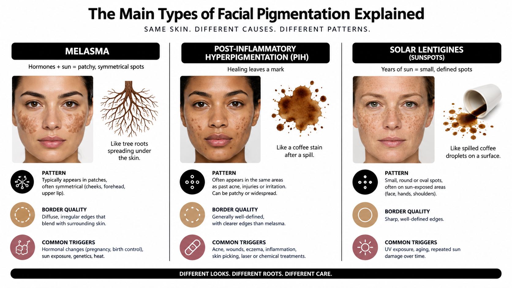
Melasma
Melasma usually appears as broader, fairly symmetrical patches on the cheeks, forehead, nose or upper lip. The edges can be irregular rather than sharply outlined, and the colour may look light brown, medium brown or grey-brown. Hormonal changes, visible light, ultraviolet exposure and heat can all influence it.
Melasma is often confused with sunspots because both can look brown. A useful comparison is a tree root system. Melasma tends to spread in connected, branching areas, while an isolated sunspot behaves more like a distinct mark. The difference between melasma and sunspots matters because a treatment that suits a discrete lesion may aggravate a diffuse, reactive pattern.
Post-inflammatory hyperpigmentation
PIH appears after the skin has been irritated or injured. A spot may follow acne, a scratch, a rash, picking, waxing, burns or a cosmetic procedure. It's usually more scattered and often corresponds to the original problem rather than following a symmetrical facial map.
Think of coffee spilled onto a tablecloth. If the cloth is smooth and the spill stays near the surface, cleaning is more straightforward. If the liquid soaks into the fibres, the stain becomes harder to lift. In skin, pigment deposited deeper after inflammation behaves less predictably and may fade slowly.
Solar lentigines
Solar lentigines, or sunspots, are usually discrete brown marks with a clearer border. They often appear on areas that have received cumulative sun exposure, such as the cheeks, temples, forehead and hands. Their appearance can overlap with freckles or other lesions, so a changing, bleeding or unusual mark needs medical assessment rather than cosmetic treatment.
The practical complication is mixed pigmentation. Melasma, PIH and sunspots may occupy the same face, but they don't share the same trigger or response. Correctly identifying the dominant type is the first major treatment decision.
How a Clinician Diagnoses Your Pigmentation
A useful consultation feels more like investigation than shopping. The clinician begins by examining distribution, symmetry, border quality, colour variation and the relationship between the marks and previous inflammation. Your history then adds context: when the pigmentation began, whether it changed during pregnancy or contraception, what products or procedures you've tried, and how your skin reacted.
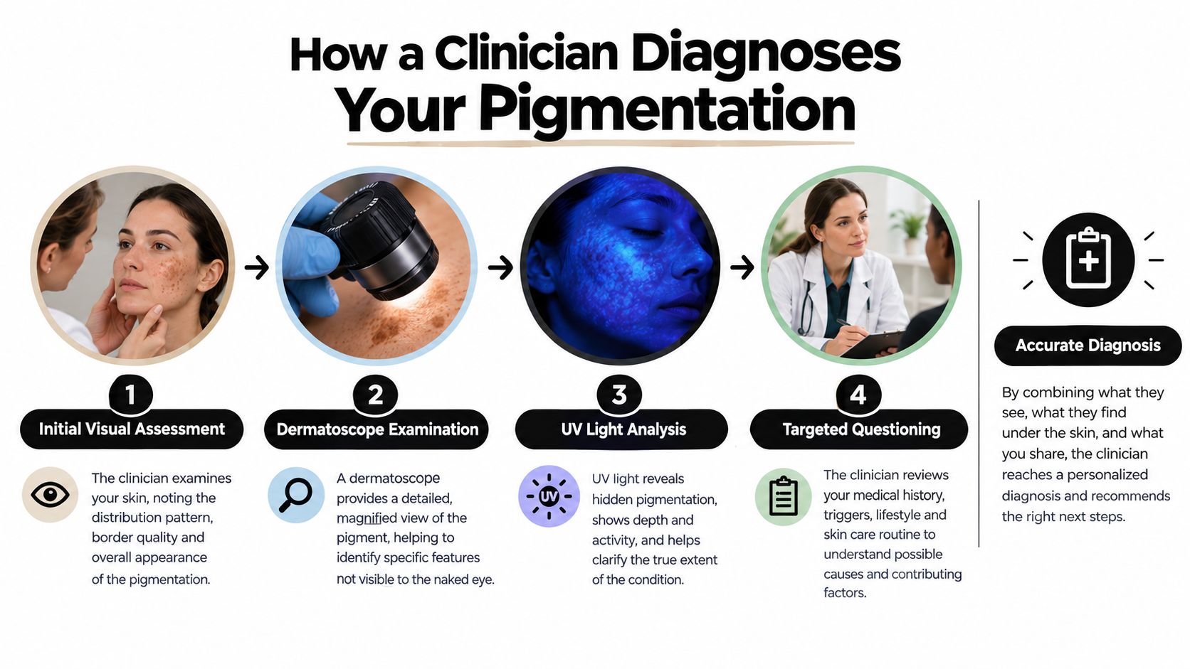
Assessing the surface and depth
A Wood's lamp can help a clinician estimate whether pigment is more apparent in the superficial layers or less clearly defined at depth. It isn't a perfect answer, but it contributes to the treatment decision. A dermatoscope can reveal structures and colour patterns that aren't obvious to the naked eye, while consistent photography provides a reliable baseline for later comparison.
Standardised lighting matters. A face photographed beside a window may appear lighter or darker depending on the time of day, camera settings and angle. A clinic that documents the skin consistently can distinguish genuine change from a flattering photograph.
Reading mixed cases carefully
When melasma sits over PIH, the clinician may first reduce inflammation and stabilise the skin rather than treating every mark immediately. If the diagnosis remains uncertain, referral or further medical assessment is more appropriate than proceeding with a high-energy procedure.
The danger of skipping this reasoning is familiar. A laser can fragment pigment in a discrete lesion yet provoke inflammation across a melasma-prone area. In darker skin, that inflammation can create new PIH, leaving the patient with a larger or more uneven problem than the one they wanted treated.
The consultation should end with a diagnosis or a clear explanation of uncertainty, not just a machine recommendation. This short clinical video offers another visual introduction to the assessment process:
Topical Treatments and What They Actually Do
Topicals work best when each ingredient has a defined role. A common mistake is adding several irritants at once, then interpreting the resulting redness as proof that the products are working. Irritation can stimulate more PIH, especially in skin already prone to inflammation.
| Ingredient | How it works | Best for | Limitations |
|---|---|---|---|
| Hydroquinone | Reduces melanin production | Short, supervised courses for stubborn epidermal pigmentation | Can irritate, requires professional guidance and may be cycled |
| Azelaic acid | Helps regulate pigment production and inflammation | PIH, acne-prone skin and some melasma patterns | Results are gradual and irritation can occur |
| Tranexamic acid | Helps reduce pathways associated with pigment signalling | Melasma and recurrent uneven tone | Oral use needs medical assessment; topical results vary |
| Retinoids | Increase cell turnover and support pigment treatment | PIH and combination routines | Dryness and irritation can worsen colour if introduced too quickly |
| Vitamin C | Provides antioxidant support and may brighten uneven tone | Maintenance and mild discolouration | Formulas vary and it won't correct every deep pigment problem |
| Niacinamide | Supports the skin barrier and may reduce transfer of pigment | Sensitive skin and maintenance | Usually needs to be part of a wider plan |
| Arbutin or kojic acid | Helps reduce melanin production | Mild or maintenance-focused routines | May be insufficient for established or deeper pigmentation |
| Broad-spectrum SPF | Limits ongoing ultraviolet stimulation | Every pigmentation type | It protects progress but doesn't erase existing pigment alone |
Hydroquinone remains a clinically important option, but it isn't a casual add-on. A clinician may use it for a defined period, monitor tolerance and then move to a maintenance routine. Prescription treatment can be more appropriate than an expanding over-the-counter shelf when melasma or deeper pigment has persisted.
Tranexamic acid also needs distinction. A topical formulation and oral treatment aren't interchangeable. Oral tranexamic acid requires a medical review because suitability depends on your health history and clotting risk.
For a plain-language overview of home routines, how to get rid of dark spots can help you understand the difference between pigment-supporting ingredients and realistic expectations. Whichever routine you choose, introduce changes gradually and judge progress over months, not days.
LED can be used as a supportive treatment in some skin plans, but it isn't a pigment-destroying substitute for diagnosis. Details about LED light therapy facials should be considered alongside, rather than instead of, a plan for the underlying pigment type.
Peels, Lasers and Light-Based Options Compared
Procedures differ in how much controlled injury they create and which pigment depth they can influence. A superficial peel may refresh the epidermis, while a laser can deliver highly concentrated energy to selected pigment. More energy isn't automatically better, especially when the skin is reactive or naturally produces more melanin after inflammation.
| Modality | Pigment depth targeted | Best suited to | Main risk |
|---|---|---|---|
| Glycolic or lactic peel | Mainly superficial epidermal pigment | Selected PIH and surface dullness | Irritation and rebound PIH |
| Salicylic peel | Surface pigment with an oil-soluble action | Acne-associated marks and oily skin | Dryness, irritation and uneven healing |
| TCA or modified Jessner peel | Deeper epidermal action, depending on strength | Carefully selected lesions and experienced hands | Excessive inflammation, scarring or PIH |
| Low-fluence Q-switched Nd:YAG | Melanin-selective pigment fragmentation | Selected melasma patterns and darker skin tones | Rebound pigmentation or mottled lightening |
| Picosecond laser | Short-pulse pigment fragmentation | Selected discrete pigment, depending on diagnosis | Inflammation and unpredictable response |
| IPL | Broad light treatment across superficial pigment and redness | Carefully selected lighter skin and sun-related lesions | Burns, PIH and poor targeting in unsuitable skin |
| Fractional non-ablative laser | Controlled dermal remodelling with limited surface injury | Texture and selected pigment patterns | Persistent inflammation or pigment change |
| Ablative fractional laser | Deeper resurfacing and remodelling | Carefully selected scars and texture problems | Greater downtime, PIH and scarring |
UK guidance specifically favours low-fluence Q-switched lasers for darker skin tones in suitable cases because they can target melanin with less epidermal heating than aggressive resurfacing approaches (UK pigmentation treatment guidance). That doesn't make the device universally safe. The diagnosis, settings, patch testing, treatment interval and aftercare still matter.
Peels can be useful when the problem is predominantly superficial, but a strong peel on active melasma may create the inflammation that drives its return. IPL can be effective for some discrete sun-related lesions, yet broad-spectrum light is less selective than a pigment-specific pulse and needs careful patient selection.
A laser may temporarily clear visible colour without changing the tendency that produced it. For melasma, the sensible approach is usually conservative sequencing, maintenance topicals and rigorous light protection. If you're considering a laser pigmentation removal treatment, ask what diagnosis the device is being used to treat, not just which machine the clinic owns.
Who Is a Poor Candidate and What Can Make It Worse
A patient can arrive with a few dark patches and still be a poor candidate for immediate treatment. Unstable, recently sun-exposed, actively inflamed or incorrectly diagnosed skin may react badly, even when the pigmentation looks mild. Removing visible colour without addressing its trigger is like repainting a damp wall. The mark may return, or inflammation may create new pigment.
Darker skin tones need careful assessment and conservative settings. UK expert sources report limited confidence and training among clinicians managing skin-of-colour conditions. These findings do not rule out treatment. They show why experience with your skin tone, pigment pattern and likely response should influence the plan.
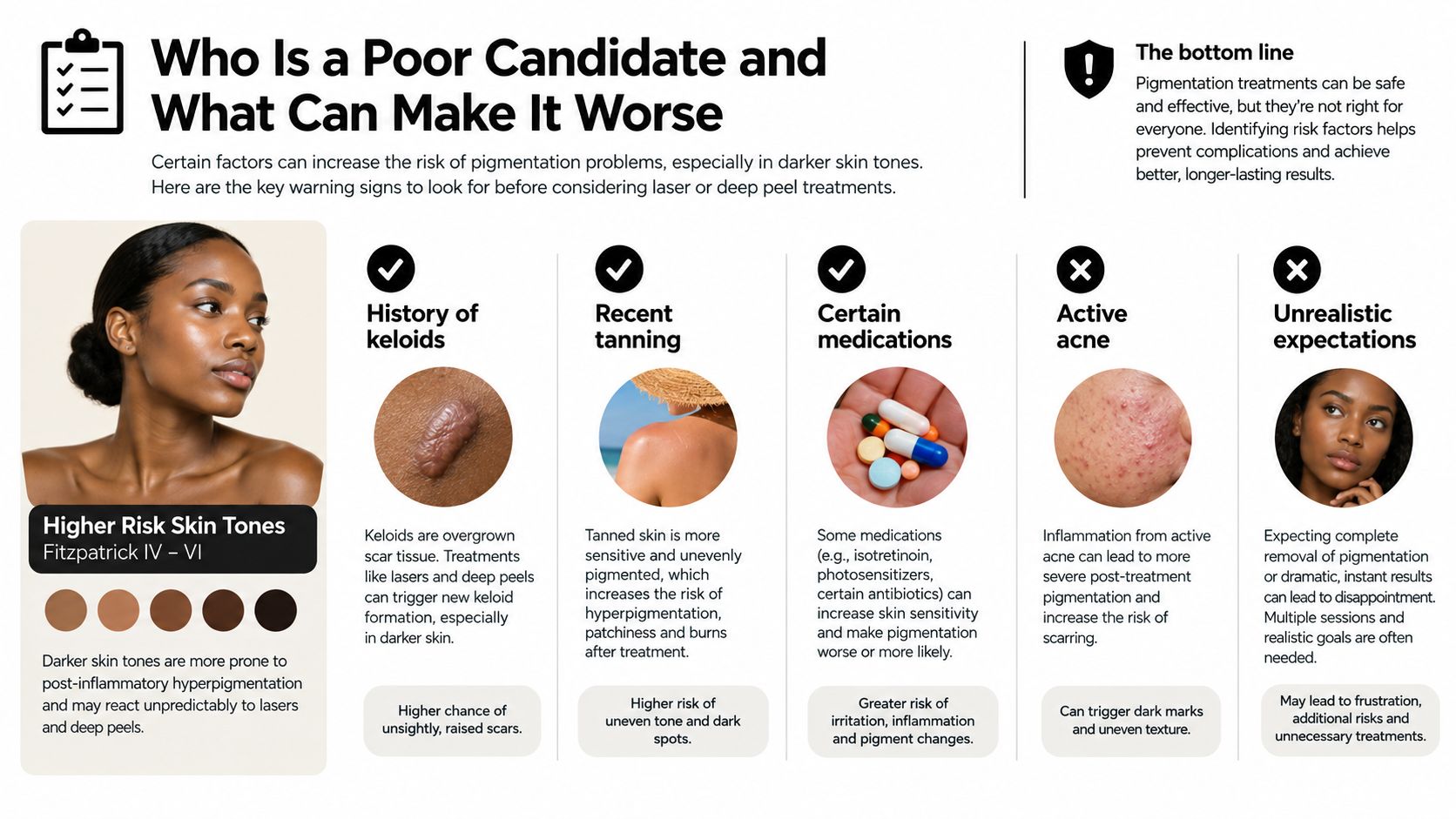
Situations that deserve caution
- Darker Fitzpatrick skin types: Aggressive lasers and deep peels can increase the risk of PIH if energy, pulse duration and recovery are not adapted.
- Recent tanning or strong sun exposure: Stimulated melanocytes may respond unpredictably to heat, light and inflammation.
- Active acne, eczema or dermatitis: Treating inflamed skin can add another pigment trigger.
- Pregnancy or changing hormonal medication: Melasma may be unstable, and some prescription ingredients or procedures may not be suitable.
- Mixed or uncertain pigmentation: A treatment can improve one pigment pattern while aggravating another.
- A history of abnormal scarring: The plan may require modification, referral or a decision not to proceed.
Exogenous ochronosis shows why a “dark patch” is not a diagnosis. Guidance on clinical guidance on melasma and facial hyperpigmentation notes that the triggering cream should be stopped and that fading can be gradual. Nd:YAG treatment remains an option, although evidence is limited.
A safer first step is sometimes no procedure. Stabilising the skin barrier, reducing inflammation and protecting against light can stop a difficult pigment problem from becoming harder.
Planning Your Treatment and Choosing the Right Clinic
A credible consultation should leave you with a written explanation, not just a package recommendation. Ask the clinician to name the suspected pigment type, explain whether it appears epidermal or dermal, and describe why the proposed treatment matches that finding.
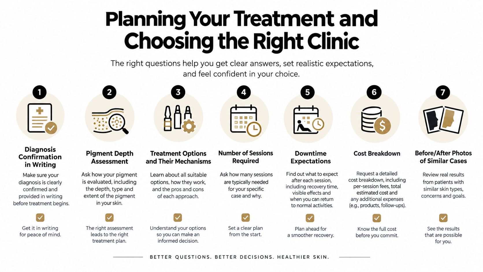
Take these questions with you:
- What is the diagnosis? Ask for it in writing, including whether the clinician suspects melasma, PIH, solar lentigines or a mixed pattern.
- How deep does the pigment appear? Ask whether a Wood's lamp, dermatoscope or other assessment supports the conclusion.
- How will my skin type affect the plan? The clinic should discuss Fitzpatrick typing, previous reactions and whether a patch test is appropriate.
- What are the alternatives? A thoughtful plan may include topicals, observation, light protection or referral instead of an immediate laser.
- What will recovery involve? Clarify redness, swelling, peeling, work restrictions and aftercare.
- How will progress be measured? Consistent photographs are more useful than casual before-and-after images.
- What happens if the pigmentation worsens? You should know who reviews complications and how the plan changes.
A clinic should also explain session planning and total cost without hiding essential aftercare or maintenance behind a later sales conversation. It's reasonable to ask to see results from people with a similar skin tone and a comparable diagnosis. General facial services vary widely, so a guide to comparing spa facial services near Roselands may help you think about what a service includes, although a standard facial isn't equivalent to medical pigment assessment.
Warning signs
Be cautious if a clinic promises complete removal, sells a one-device-fits-all package, skips diagnosis, gives minimal sun-protection advice or pressures you to book immediately. Another warning sign is stacking aggressive treatments without explaining how cumulative inflammation could affect PIH.
A careful practitioner may decline to laser unstable melasma and suggest a period of topical preparation first. That restraint is a sign of judgement, not a lack of treatment options.
Realistic Results, Maintenance and What Comes Next
Results depend on the diagnosis you started with. Melasma is managed rather than cured, and it can recur when hormonal, light or heat-related triggers remain active. PIH often fades substantially but slowly, particularly when the original inflammation continues. Discrete sun-related lesions may respond more directly to a suitable light-based treatment, but new lesions can still develop with ongoing exposure.
The NHS describes an important distinction in another pigment condition. In its February 2026 announcement, NHS England said nearly 100,000 people aged 12 and over could be offered ruxolitinib cream for non-segmental vitiligo, including facial repigmentation, and reported that clinical trials showed many patients achieved at least a 75% return of skin colour to affected areas, with results usually reviewed after about six months (UK aesthetics industry reference covering the NHS announcement). Vitiligo is not hyperpigmentation, but the development illustrates how carefully diagnosed pigment disorders can require structured, long-term treatment rather than a quick cosmetic fix.
Maintenance may include a prescribed pigment inhibitor, a gentler brightening routine, daily broad-spectrum SPF and avoidance of known triggers such as friction, excess heat or unsuitable hormonal products. Some people also need occasional top-up treatment, while others do better by maintaining a stable home routine and avoiding unnecessary procedures.
The goal is control with the least irritation needed to achieve it.
Many patients feel more comfortable socially when a persistent patch becomes less visible, but confidence usually follows steady improvement rather than one dramatic session. The most reliable progress comes from correct diagnosis, conservative sequencing, consistent home care and a clinician who remains involved after the procedure.
At 3D Aesthetics Leamington Spa, facial pigmentation consultations can explore options such as 3D Nanosure pigmentation treatment and the HydrO2 Pigmentation Facial according to your skin and diagnosis. Visit 3D Aesthetics Leamington Spa to discuss your pigmentation concerns, treatment suitability, aftercare and a plan focused on safe, measured improvement.
← Back to Home